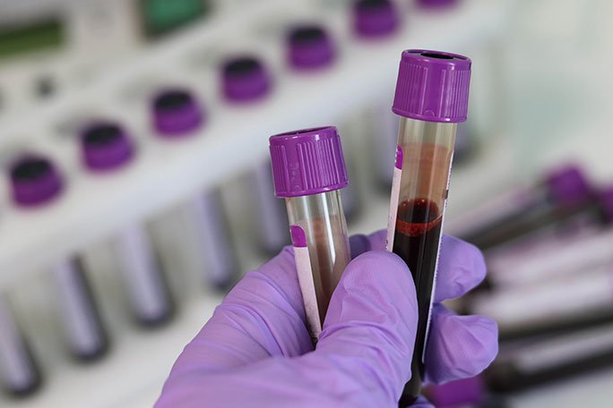
Proper Protocol for Collecting Blood Samples into a Dark Blue Top Tube
Summary
- The Dark Blue Top tube is used for certain specialized tests that require clotting to occur before centrifugation.
- Proper technique and handling of the Dark Blue Top tube are crucial to ensure accurate Test Results.
- Following the appropriate protocol during blood sample collection into a Dark Blue Top tube is essential for the success of phlebotomy procedures.
Introduction
Collecting blood samples into different colored tubes is a common practice in healthcare settings, especially during phlebotomy procedures. Each tube color corresponds to specific tests and requirements for proper specimen collection and processing. In this article, we will focus on the appropriate protocol for collecting blood samples into a Dark Blue Top tube during phlebotomy procedures in the United States.
Understanding the Dark Blue Top Tube
The Dark Blue Top tube, also known as the citrate tube, is used for specialized coagulation tests that require the blood to clot before centrifugation. These tests measure various aspects of a patient's blood clotting process and are essential for diagnosing and monitoring conditions such as bleeding disorders, Clotting Disorders, and Anticoagulant therapy effectiveness.
Indications for Use
The Dark Blue Top tube is typically used for the following coagulation tests:
- PT (Prothrombin Time)
- INR (International Normalized Ratio)
- APTT (Activated Partial Thromboplastin Time)
- Fibrinogen
Precautions
When using a Dark Blue Top tube for blood sample collection, it is important to:
- Avoid hemolysis, as it can affect Test Results.
- Fill the tube to the proper volume to ensure the correct blood-to-Anticoagulant ratio.
- Mix the blood gently and thoroughly with the Anticoagulant to prevent clot formation.
Protocol for Collecting Blood Samples into a Dark Blue Top Tube
Following the appropriate protocol during blood sample collection into a Dark Blue Top tube is crucial for obtaining accurate Test Results. Here are the steps to follow:
1. Patient Identification
Verify the patient's identity using at least two unique identifiers, such as their full name and date of birth. This ensures that the blood sample is correctly labeled and matched to the right patient.
2. Selecting the Site and Performing Hand Hygiene
Choose a suitable Venipuncture site and perform Hand Hygiene to prevent the risk of infection. Proper Hand Hygiene includes washing hands with soap and water or using an alcohol-based hand sanitizer.
3. Assembling Equipment
Gather all the necessary equipment, including the Dark Blue Top tube, needle, tourniquet, alcohol swabs, and gauze pads. Ensure that the equipment is sterile and in good condition.
4. Tourniquet Application
Apply the Tourniquet above the Venipuncture site to make the veins more visible and accessible. Avoid leaving the Tourniquet on for an extended period to prevent venous stasis and hemoconcentration.
5. Vein Selection and Needle Insertion
Select a suitable vein for Venipuncture and insert the needle at the correct angle and depth. As soon as blood enters the needle, release the Tourniquet to prevent backflow of tissue fluid into the blood sample.
6. Blood Collection into the Dark Blue Top Tube
Fill the Dark Blue Top tube with the appropriate volume of blood according to the lab's requirements. Invert the tube gently 3-4 times to mix the blood with the Anticoagulant uniformly.
7. Needle Removal and Hemostasis
After filling the Dark Blue Top tube, remove the needle from the vein and apply gentle pressure to the Venipuncture site with a sterile gauze pad to promote hemostasis. Discard the used needle into a sharps container.
8. Labeling and Transport
Label the Dark Blue Top tube with the patient's information, date and time of collection, and your initials. Ensure that the specimen is transported to the lab promptly and in the appropriate conditions to preserve Sample Integrity.
Conclusion
Proper technique and handling of the Dark Blue Top tube during blood sample collection are essential for obtaining accurate coagulation Test Results. By following the appropriate protocol outlined in this article, healthcare professionals can ensure the success of phlebotomy procedures and the delivery of quality patient care.

Disclaimer: The content provided on this blog is for informational purposes only, reflecting the personal opinions and insights of the author(s) on the topics. The information provided should not be used for diagnosing or treating a health problem or disease, and those seeking personal medical advice should consult with a licensed physician. Always seek the advice of your doctor or other qualified health provider regarding a medical condition. Never disregard professional medical advice or delay in seeking it because of something you have read on this website. If you think you may have a medical emergency, call 911 or go to the nearest emergency room immediately. No physician-patient relationship is created by this web site or its use. No contributors to this web site make any representations, express or implied, with respect to the information provided herein or to its use. While we strive to share accurate and up-to-date information, we cannot guarantee the completeness, reliability, or accuracy of the content. The blog may also include links to external websites and resources for the convenience of our readers. Please note that linking to other sites does not imply endorsement of their content, practices, or services by us. Readers should use their discretion and judgment while exploring any external links and resources mentioned on this blog.
