
Understanding the Anatomic Pathology Workflow in Clinical Labs
Anatomic pathology is a crucial component of the healthcare system, providing essential information for the diagnosis, treatment, and management of diseases. This branch of pathology involves the examination of tissue samples from patients to determine the presence and extent of disease. In clinical labs, anatomic pathology Workflow plays a significant role in ensuring accurate and timely results for patient care.
Specimen Collection and Transport
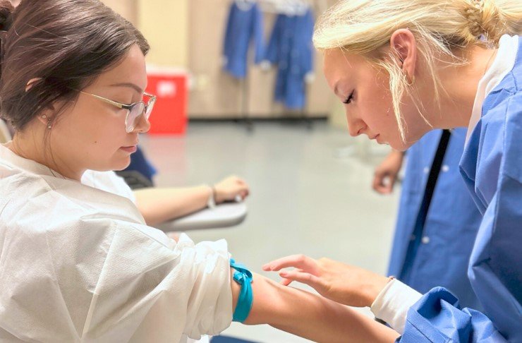
The first step in the anatomic pathology Workflow is the collection of tissue specimens from patients. This may involve biopsies, surgical resections, or autopsy samples. It is essential to ensure that the specimen is properly labeled and accompanied by relevant clinical information to guide the pathologist in their evaluation.
Once the specimen is collected, it needs to be transported to the Pathology Lab promptly and in optimal conditions to prevent degradation and ensure accurate results. Proper packaging, handling, and transportation methods are essential to maintain the integrity of the specimen.
Specimen Processing
Upon arrival at the Pathology Lab, the tissue specimen undergoes processing to prepare it for examination under the microscope. This may involve fixation, embedding, sectioning, staining, and mounting of the tissue sample on glass slides. Each step in the processing is critical to ensure that the cellular and tissue structures are preserved and visible for analysis.
Fixation
Fixation is the process of preserving the tissue specimen by immersing it in a chemical solution, usually formalin. This step prevents degradation of cellular structures and helps maintain the morphology of the tissue for microscopic examination.
Embedding
After fixation, the tissue specimen is embedded in a paraffin block to provide support for thin sectioning. The paraffin block is then sliced into thin sections using a microtome, and the tissue sections are mounted onto glass slides for staining.
Staining
Staining is a critical step in the anatomic pathology Workflow, as it helps highlight specific cellular structures and components for analysis. Different stains are used to differentiate between various cell types, detect abnormalities, and identify specific features indicative of disease.
Microscopic Examination
Once the tissue sections are prepared and stained, they are ready for microscopic examination by a pathologist. The pathologist carefully observes the cellular and tissue structures under the microscope to identify any abnormalities, lesions, or pathological changes indicative of disease.
The pathologist may employ various techniques, such as immunohistochemistry, fluorescence in situ hybridization, or molecular testing, to further characterize the tissue sample and provide a more precise diagnosis.
Diagnosis and Reporting
Based on the findings from the microscopic examination, the pathologist formulates a diagnosis and prepares a detailed report documenting their observations. The pathology report includes relevant clinical information, microscopic description of the tissue sample, diagnostic interpretation, and any additional tests or recommendations for further evaluation.
Quality Assurance and Quality Control
Quality assurance and Quality Control are essential components of the anatomic pathology Workflow in clinical labs to ensure accurate and reliable results. Pathology labs adhere to strict Quality Standards and undergo regular inspections and audits to maintain compliance with regulatory requirements.
Internal Quality Control
Internal Quality Control involves monitoring and verifying the accuracy and precision of laboratory tests and procedures. This may include routine checks of equipment, reagent performance, and Proficiency Testing to ensure consistent and reliable results.
External Quality Assurance
External quality assurance programs involve participation in Proficiency Testing schemes and external quality assessment programs to benchmark performance against other labs and ensure the accuracy and reliability of results. This helps identify any potential areas for improvement and maintain high standards of quality in anatomic pathology practice.
Technological Advancements in Anatomic Pathology
Advancements in technology have revolutionized the field of anatomic pathology, leading to greater accuracy, efficiency, and precision in diagnostic testing. Digital pathology, whole slide imaging, Artificial Intelligence, and molecular testing are some of the innovative technologies that have transformed the practice of anatomic pathology.
Digital Pathology
Digital pathology involves the digitization of glass slides and microscopic images, allowing pathologists to view and analyze tissue samples on a computer screen. This technology enables remote viewing, sharing, and consultation of digital slides, improving Workflow efficiency and collaboration among pathologists.
Whole Slide Imaging
Whole slide imaging captures high-resolution images of entire tissue sections, providing a comprehensive view of the sample for analysis. Pathologists can navigate through the digital slides, zoom in on specific areas of interest, and annotate findings, enhancing diagnostic capabilities and facilitating telepathology consultations.
Artificial Intelligence
Artificial Intelligence algorithms have been developed to assist pathologists in analyzing and interpreting complex histological images. These AI tools can aid in the detection of abnormalities, classification of tumors, and prediction of patient outcomes, enhancing diagnostic accuracy and efficiency in anatomic pathology practice.
Molecular Testing
Molecular testing involves the analysis of DNA, RNA, and protein markers in tissue samples to identify specific genetic mutations, gene rearrangements, or Biomarkers indicative of disease. This molecular information provides valuable insights into the underlying mechanisms of disease, guiding treatment decisions and Personalized Medicine approaches.
Challenges and Future Directions
Despite the advancements in anatomic pathology Workflow, there are several challenges that pathologists and clinical labs face, such as increasing workload, workforce shortages, and evolving regulatory requirements. As the demand for diagnostic testing continues to grow, pathologists must adapt to new technologies, implement efficient workflows, and collaborate with multidisciplinary teams to deliver high-quality patient care.
Looking ahead, the future of anatomic pathology holds promising opportunities for innovation and improvement in diagnostic testing, Personalized Medicine, and precision healthcare. By leveraging technology, embracing quality assurance practices, and staying abreast of emerging trends, pathologists can navigate the complexities of the healthcare landscape and continue to play a vital role in the diagnosis and management of diseases.
Related Videos
Disclaimer: The content provided on this blog is for informational purposes only, reflecting the personal opinions and insights of the author(s) on phlebotomy practices and healthcare. The information provided should not be used for diagnosing or treating a health problem or disease, and those seeking personal medical advice should consult with a licensed physician. Always seek the advice of your doctor or other qualified health provider regarding a medical condition. Never disregard professional medical advice or delay in seeking it because of something you have read on this website. If you think you may have a medical emergency, call 911 or go to the nearest emergency room immediately. No physician-patient relationship is created by this web site or its use. No contributors to this web site make any representations, express or implied, with respect to the information provided herein or to its use. While we strive to share accurate and up-to-date information, we cannot guarantee the completeness, reliability, or accuracy of the content. The blog may also include links to external websites and resources for the convenience of our readers. Please note that linking to other sites does not imply endorsement of their content, practices, or services by us. Readers should use their discretion and judgment while exploring any external links and resources mentioned on this blog.